Anatomy Of Chest : Anatomy Thoracic Cavity - The chest wall is comprised of skin, fat, muscles, and the thoracic skeleton.
byAdmin•
0
Anatomy Of Chest : Anatomy Thoracic Cavity - The chest wall is comprised of skin, fat, muscles, and the thoracic skeleton.. This page provides an overview of the chest muscle group. Here, we break down the anatomy of your chest muscles. It spreads out like a fan and covers the rib cage like an armor plate. The muscles of the chest develop from the somites found in the mesoderm. The epidermis is the outermost layer that provides a protective, waterproof seal over the body.
Learn about each of these muscles, their locations, functional anatomy and exercises for them. 12 photos of the anatomy of the chest and stomach. Sternocleidomastoid muscle clavicle and ribs anatomy muscle anatomy chest sternocleidomastoid ribs anatomy chest muscles anatomy thorax rib muscles chest muscles chest anatomy illustration. See chest anatomy stock video clips. It spreads out like a fan and covers the rib cage like an armor plate.
Human Chest Anatomy Illustration Stock Image F025 1029 Science Photo Library from media.sciencephoto.com The human thorax includes the thoracic cavity and the thoracic wall. 12 cm (5 in) in length, 8 cm (3.5 in) wide, and 6 cm (2.5 in) in thickness. The myotomes elongate and invade the mesoderm of the wall of the embryonic thoracic and abdominal cavities. System respiratory respiratory organs of human body digestive and respiratory system medical chest internal structure of human body medicine body lungs biology intestines stomach anatomy torso human internal. Three dimensional view of the female reproductive system, full frontal view. Principal functions are the protection of internal viscera and an expandable cylinder facilitating variable gas flow into the lungs. The thorax has two major openings: Anatomy of right side chest pain.
Thoracic cavity, also called chest cavity, the second largest hollow space of the body.
31 anatomy of the female breast syllabus p. The superior thoracic aperture found superiorly and the inferior thoracic aperture. The chest is made up primarily of two muscles: The epidermis is the outermost layer that provides a protective, waterproof seal over the body. System respiratory respiratory organs of human body digestive and respiratory system medical chest internal structure of human body medicine body lungs biology intestines stomach anatomy torso human internal. Knowledge of the anatomy of the whole cylinder (ribs, sternum, vertebra, diap … It provides protection to vital organs (eg, heart and major vessels, lungs, liver) and provides stability for movement. The thorax has two major openings: A good radiologist knows the anatomy because knowing where structures normally live and recognizing the location of an abnormality helps to make or narrow the differential diagnosis. The chest anatomy includes the pectoralis major, pectoralis minor and the serratus anterior. Thoracic cavity, also called chest cavity, the second largest hollow space of the body. 12 cm (5 in) in length, 8 cm (3.5 in) wide, and 6 cm (2.5 in) in thickness. Swensen fund for innovation in teaching.
The thorax has two major openings: The epidermis is the outermost layer that provides a protective, waterproof seal over the body. Knowledge of the anatomy of the whole cylinder (ribs, sternum, vertebra, diap … Radiology basics of chest ct anatomy with annotated coronal images and scrollable axial images to help medical students and junior doctors learning anatomy. Principal functions are the protection of internal viscera and an expandable cylinder facilitating variable gas flow into the lungs.
Chest Wall Thoracic Cavity And Pleura Sanjaya Adikari Department Of Anatomy Ppt Download from images.slideplayer.com 12 photos of the anatomy of the chest and stomach. The chest wall, like other regional anatomy, is a remarkable fusion of form and function. 2 skin of the anterior chest wall syllabus p. Anatomy of the thorax, heart, abdomen and pelvis recommended text gray's anatomy for students. Anatomy of right side chest pain. Related posts of anatomy of the chest area anatomy of the female human body. 4 innervation of the breast blood supply of the breast syllabus p. The upper part of your pec major, the clavicular head runs from your clavicle (collarbone) across the top of your chest and attaches to your humerus, or upper arm.
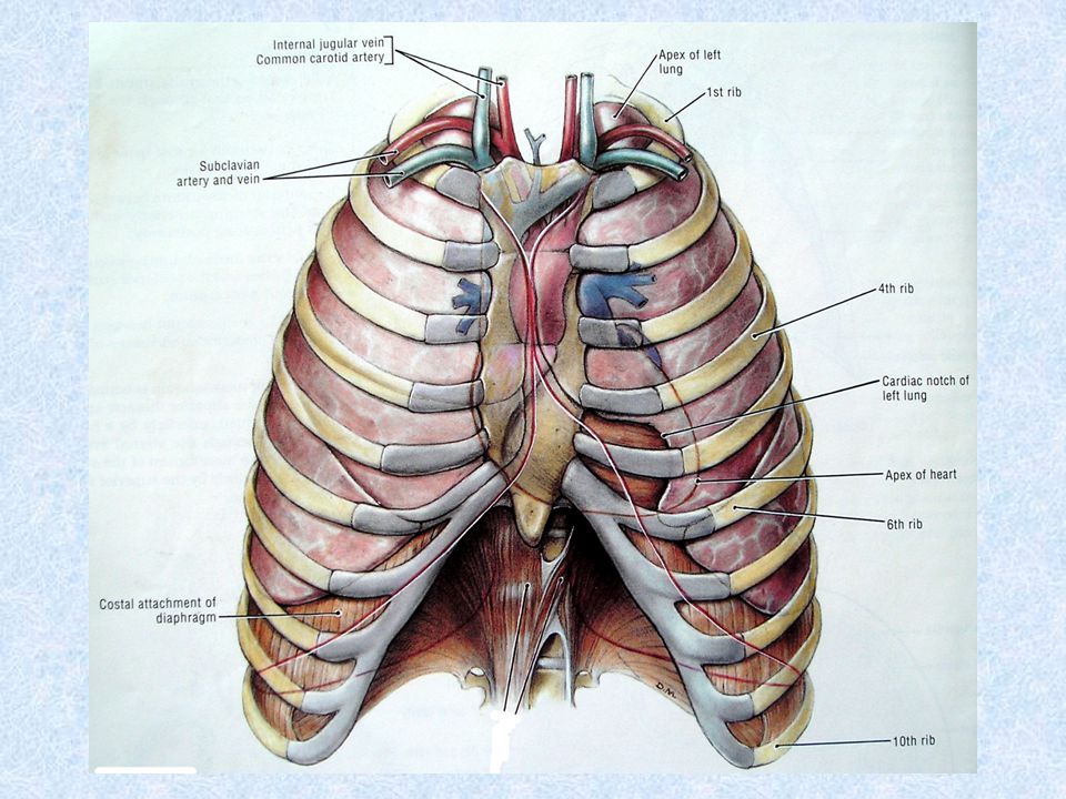
A line is drawn from anterior surface of the body of 6th thoracic vertebrae passing through the apex of the heart up to anterior lower most part of diaphragm.
Anatomically, the heart is located in the anterior thoracic cavity; Anatomy of right side chest pain. The chest anatomy includes the pectoralis major, pectoralis minor and the serratus anterior. Browse 6,407 chest anatomy stock photos and images available, or search for human anatomy to find more great stock photos and pictures. The chest or thorax is the region between the neck and diaphragm that encloses organs, such as the heart, lungs, esophagus, trachea, and thoracic diaphragm. These myotomes divide into the epimere and the hypomere. Radiology basics of chest ct anatomy with annotated coronal images and scrollable axial images to help medical students and junior doctors learning anatomy. The epidermis is the outermost layer that provides a protective, waterproof seal over the body. This mri chest (thorax) axial cross sectional anatomy tool is absolutely free to use. Plus, how to target each to make them bigger and stronger. A good radiologist knows the anatomy because knowing where structures normally live and recognizing the location of an abnormality helps to make or narrow the differential diagnosis. See human chest anatomy stock video clips. A line is drawn from anterior surface of the body of 6th thoracic vertebrae passing through the apex of the heart up to anterior lower most part of diaphragm.
Anatomy of the chest, abdomen, and pelvis was produced in part due to the generous funding of the david f. This mri chest (thorax) axial cross sectional anatomy tool is absolutely free to use. Swensen fund for innovation in teaching. A typical heart is approximately the size of your fist: It provides protection to vital organs (eg, heart and major vessels, lungs, liver) and provides stability for movement.
Venous Chest Anatomy Clinical Implications Sciencedirect from ars.els-cdn.com The chest wall is comprised of skin, fat, muscles, and the thoracic skeleton. Anatomy of the thorax, heart, abdomen and pelvis recommended text gray's anatomy for students. Anatomy of right side chest pain. Computed tomography (ct) of the chest can detect pathology that may not show up on a conventional chest radiograph(1). About the 6th week, the somites differentiate into the sclerotomes and the dermatomyotomes. Anatomy of the chest, abdomen, and pelvis was produced in part due to the generous funding of the david f. It is enclosed by the ribs, the vertebral column, and the sternum, or breastbone, and is separated from the abdominal cavity (the body's largest hollow space) by a muscular and membranous partition, the diaphragm. Anatomically, the heart is located in the anterior thoracic cavity;
12 cm (5 in) in length, 8 cm (3.5 in) wide, and 6 cm (2.5 in) in thickness.
Thoracic cavity, also called chest cavity, the second largest hollow space of the body. Use the mouse scroll wheel to move the images up and down alternatively use the tiny arrows (>>) on both side of the image to move the images.>>) on both side of the image to move the images. The muscles of the chest develop from the somites found in the mesoderm. 2 skin of the anterior chest wall syllabus p. Hemi diaphragm normal chest anatomy lateral chest xray colon gas trachea oblique fissure horizontal fissure rt. System respiratory respiratory organs of human body digestive and respiratory system medical chest internal structure of human body medicine body lungs biology intestines stomach anatomy torso human internal. (1) the pectoralis major, and (2) the pectoralis minor. Computed tomography (ct) of the chest can detect pathology that may not show up on a conventional chest radiograph(1). Browse 6,407 chest anatomy stock photos and images available, or search for human anatomy to find more great stock photos and pictures. Radiology basics of chest ct anatomy with annotated coronal images and scrollable axial images to help medical students and junior doctors learning anatomy. A good radiologist knows the anatomy because knowing where structures normally live and recognizing the location of an abnormality helps to make or narrow the differential diagnosis. The chest or thorax is the region between the neck and diaphragm that encloses organs, such as the heart, lungs, esophagus, trachea, and thoracic diaphragm. Anatomy of the thorax, heart, abdomen and pelvis recommended text gray's anatomy for students.